Cryogenic electron microcopy (cryo-EM) is an advanced imaging technique that is used to determine the three-dimensional structure of biomolecules after rapidly freezing and trapping them in vitreous ice. It allows researchers to visualize complex biological assemblies without the need for crystallization. CryoEM is an indispensable tool for structural biologists, it aids structure elucidation of single molecules, biomolecular complexes, viruses, and cells.
Cryo-EM equipment is expensive and requires expert users to operate. In addition, sample preparation for cryo-EM is a complex, tedious process that requires a certain level of sample homogeneity to maximize resolution. Producing high-quality samples that are uniform and free of contaminants can be difficult. While cryo-EM can achieve near-atomic resolution, the quality of sample plays a huge role in achieving the highest possible resolution.
Ensuring samples meet a minimum quality threshold, before vitrification, is advantageous in streamlining the cryo-EM workflow.
Negative Staining
Negative staining is an established technique used in electron microscopy to enhance the contrast of biological specimens. It involves surrounding the sample with a dense, electron-opaque stain that does not penetrate the specimen but instead fills the spaces around it. This creates a high-contrast image where the specimen appears light against a dark background, allowing for better visualization of its structure. Negative staining is used extensively to visualize the morphology of viruses, aiding in the identification and classification of viral particles, and observing the overall shape of single protein molecules and assembly of large biomolecular complexes.
It also serves as a preliminary step before more detailed structural studies, such as cryo-EM, providing initial insights into the sample. During this step, researchers can quickly check the protein size, shape, concentration, and aggregation state at room temperature. This triaging step ensures that the samples meet a minimum quality threshold and reduces the number of samples that are vitrified and imaged by cryo-EM, making the overall workflow much more efficient.
Many laboratories use a transmission electron microscope (TEM) or a scanning transmission electron microscope (STEM) to image the negatively stained samples. These instruments are expensive and require specially trained users to operate them. Using these expensive instruments to check for quality of the samples is underutilizing their capabilities and can be significant strain on the resources. Several laboratories and core facilities use older TEMs that are close to their end-of-life to image the negative stained samples. While these microscopes provide high-resolution imaging, they’re often well beyond their serviceable life. Other research groups have abandoned negative staining because supporting an additional standalone TEM in the laboratory involves significant space, personnel, service, and utilities costs. Research groups could outsource negative stain imaging, but this would drastically slow down sample optimization.
“The Pharos’ STEM feature is much simpler, faster, and more stable than our in-house TEM and would greatly speed up iterative experimentation involving negative stain grids produced.”
– Chase Budell, Staff Scientist, NYSBC –
Phenom Pharos Desktop STEM
The Phenom Pharos is a state-of-the-art desktop scanning electron microscope (SEM) that features a field emission gun (FEG) source. It provides exceptional image clarity and detail, allowing for the visualization of nanoscale structures with resolution down to the nanometer level. The Phenom Pharos is equipped with an intuitive software interface and automated functions, making it suitable for both experienced researchers and novice users. Combined with the Phenom STEM detector, the platform provides an affordable and simple-to-operate solutions for cryo-EM groups and core facilities either to replace aging room-temperature TEMs or to start using negative staining as a screening tool. The Pharos STEM detector features a segmented electron detector capable of producing bright field (BF), dark field (DF), and high-angle annular dark field (HAADF) STEM images. Integrated into a sample holder that accommodates standard 3-mm sample grids, accessing STEM mode involves the familiar action of exchanging specimens.
“The Phenom is a cost-effective, fast and easy way to detemine sample homogeneity using negative staining approach.”
– Daija Bobe, Staff Scientist, NYSBC –
Put the Phenom Nearly Anywhere in Your Lab
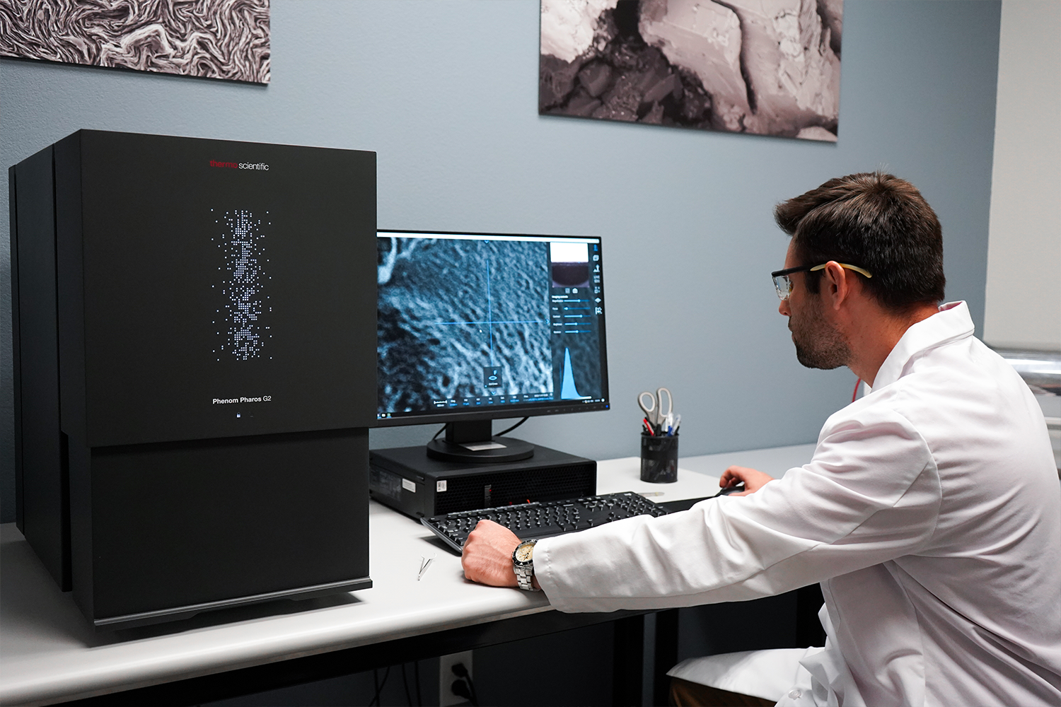
The Phenom Pharos is a compact SEM, that can be placed nearly anywhere in a TEM facility (Figure 1). This can be close to a wet bench, in the protein purification lab, or wherever it’s needed most. It fits where TEM’s cannot and does not require special power hookups, vibration isolation, field cancellation, gas lines, tension tanks, large external pumps, or chillers. The Phenom Pharos requires about three feet of bench space, a 110V outlet, and space to put a football sized roughing pump on the floor.
Figure 1. Phenom Pharos Desktop SEM in use on a laboratory benchtop.
Exceptional Throughput and Accessibility for All Users
The time it takes from loading the sample into a Phenom instrument to start acquisition of high-magnification images can be under a minute. Basic training required for an inexperienced user to operate the microscope can be done within an hour. This makes the Phenom extremely fast and convenient for any user in the laboratory to image negatively stained samples.
Affordability
The Phenom is far more affordable to own and operate than a full-sized TEM or STEM. The Phenom is approximately less than a third the cost of a LaB6 room temperature TEM. It also doesn’t require any facilities modifications or hiring of additional personnel. The service contract and ongoing operating costs are also much lower, making the Phenom more cost effective to both acquire and maintain.
Improved Productivity and Expanded Capabilities
The Phenom allows cryo-EM labs to replace an aging TEM, offload TEM or STEM used for negative stain imaging, or add negative stain imaging for the first time. With Phenom Pharos, researchers can use Phenom SEM solely for negative stain imaging, helping maximize productivity and reduce costs in cryo-EM workflow. Beyond STEM, Phenom Pharos can expand the possibilities of sample analysis by delivering high-resolution SEM imaging and energy-dispersive X-ray spectroscopy (EDS) with results on par with floor-model instruments.
Screening Negative Stained Samples
The goal of imaging negative stained samples is to confirm that the sample quality is good enough to move into cryo-EM sample preparation. Phenom Pharos yields images that are sufficient for structural biologists to make assessments of particle size, shape, aggregation status, and concentration. The resolution of the Pharos is sufficient to determine pass/fail status for many samples.
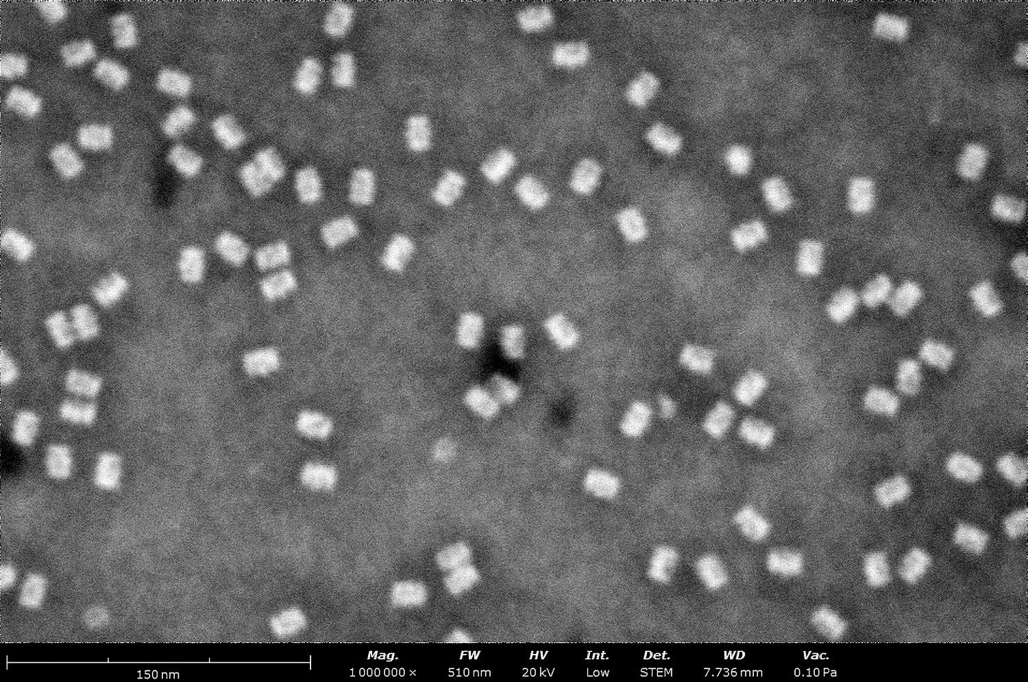
STEM image of negative stained proteasomes. The proteasome is a multi-protein complex that degrades misfolded, damaged, or unneeded proteins into peptides, playing a critical role in cellular protein homeostasis. (Sample courtesy of NYSBC.)
STEM image of negative-stained encapsulin particles. Encapsulin is a protein nanocompartment found in bacteria and archaea, used to encapsulate enzymes or cargo proteins for metabolic or protective functions.
Figure 2. STEM micrograph of negatively stained (Left) 20s proteosome, (Right) Encapsulin particles obtained on a Phenom Pharos.
The Phenom Pharos underwent rigorous testing at NY Structural Biology Center to determine whether it’s a suitable replacement room-temperature TEM for screening negative stained samples. During this evaluation, five negatively stained samples were imaged: 20s proteosome (Figure 2A), horse spleen apoferritin, bovine thyroglobulin, Myxococcus xanthus encapsulin (Figure 2B), and PP7-phage virus-like particles (Figure 3).
A BF-STEM micrograph (Figure 2) from the negatively stained 20s proteosome and encapsulin acquired on the Phenom Pharos clearly shows that the particles are of the expected shape and size and that the sample is monodisperse. Little to no aggregation was observed thus marking this preparation as “good enough” for subsequent cryo-EM sample preparation.
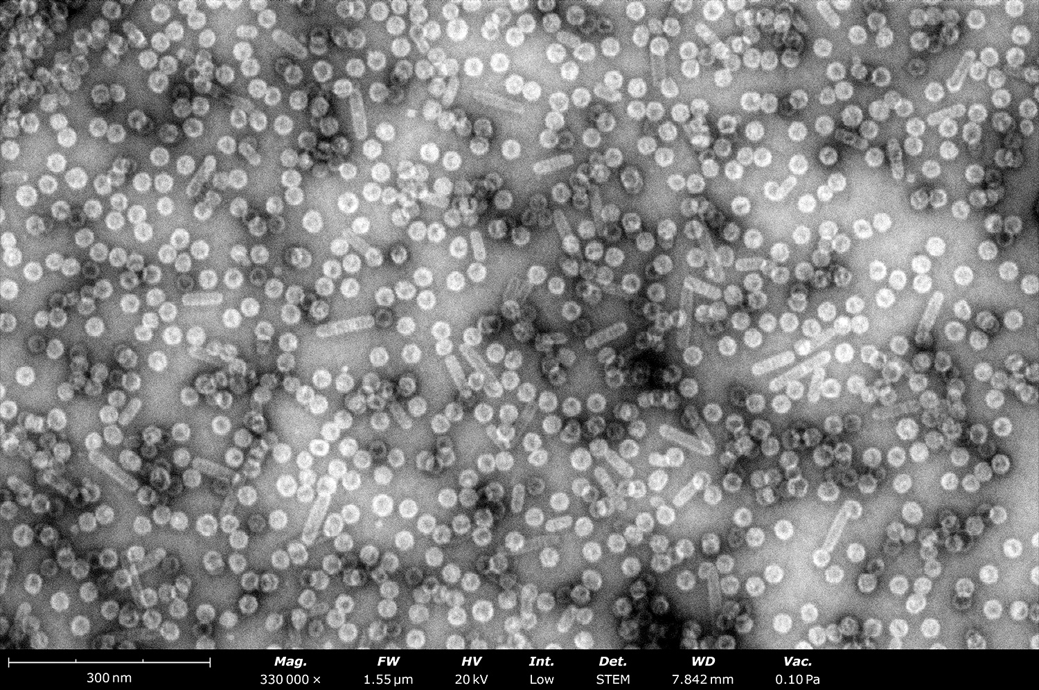
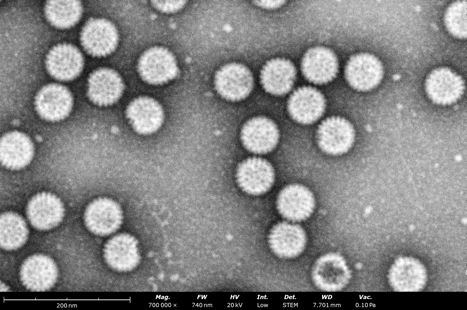
STEM image of negative-stained virus like particles. The image shows two types of VLPs: circular-shaped particles that are approximately 25 nm in size and rod-shaped particle of various lengths. Virus-like particles are self-assembled, non-infectious protein structures that mimic the organization of viral capsids and are commonly used in vaccine development and drug delivery. (Sample courtesy of NYSBC.)
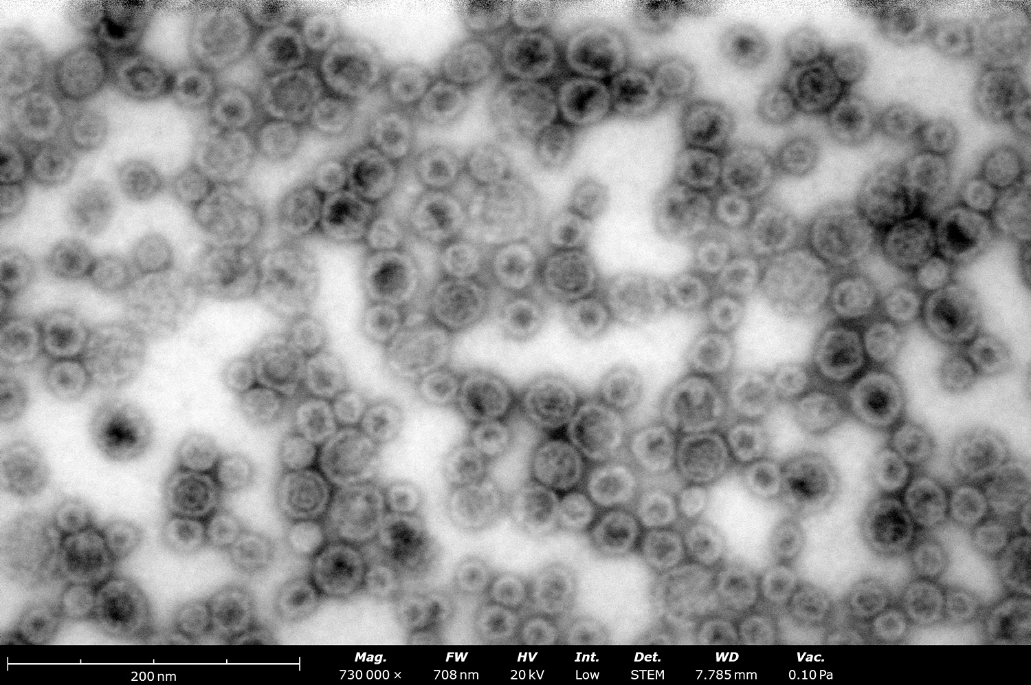
STEM image of negative-stained rotavirus particles. Rotavirus is a highly contagious double-stranded RNA virus that causes severe gastroenteritis, particularly in infants and young children, leading to significant morbidity and mortality worldwide. (Sample courtesy of Penn State University.)
Figure 3. High magnification STEM image of PP7 virus-like particles (Left) obtained on a Phenom Pharos, showing icosahedral as well as tubular assemblies adopted by these VLPs. Image of Rotavirus (Right) with VP4 projections visible (courtesy of the Kelly Lab, Penn State University).
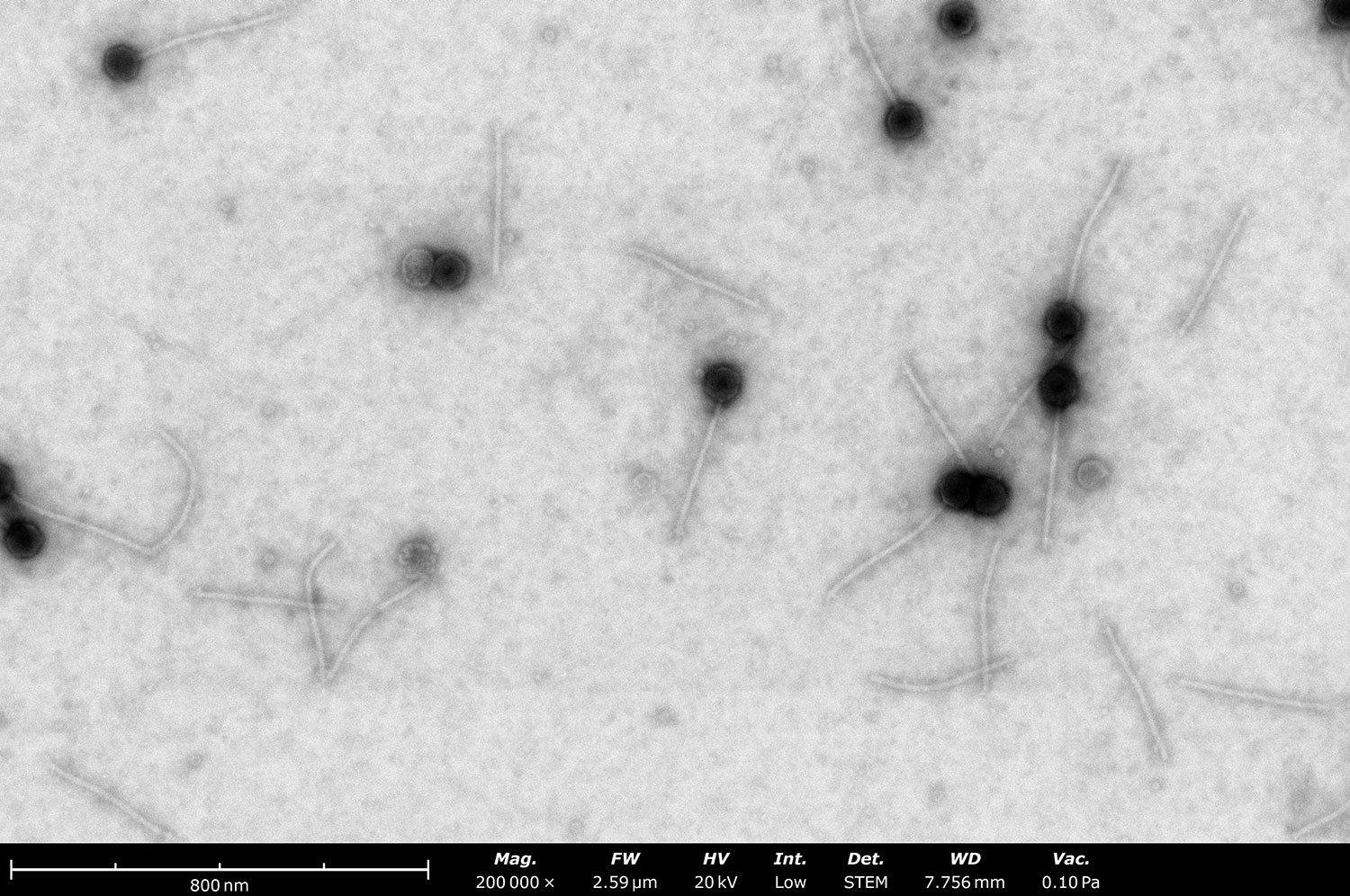
Phage Imaging
Negative staining is also an important stand-alone technique for imaging viruses and bacteriophages. For classification of bacteriophages into families it is often important to know the head size and shape and the tail length. Phage groups often rely on sequencing alone since room temperature TEMs are often inaccessible, and cryo-EM is expensive. For laboratories that study virus-like particles and their modification, imaging is a critical step to check the assembly state of their particles. The Phenom Pharos yields valuable information about phage samples without the need for an in-house TEM, special facilities, or a trained microscopist (Figure 4).
“Phenom is good enough for 90% of the negative stain imaging we do in our lab.”
– Dr. Misha Kopylov, Staff Scientist, NYSBC –
Figure 4. BF-STEM image of negatively stained long-tailed bacteriophage sample (courtesy of NYSBC) acquired on the Phenom Pharos. This image confirms that this species of bacteriophage has a large ~90 nm icosahedral head and very long flexible tails.
Phenom Pharos SEM for Grid Inspection
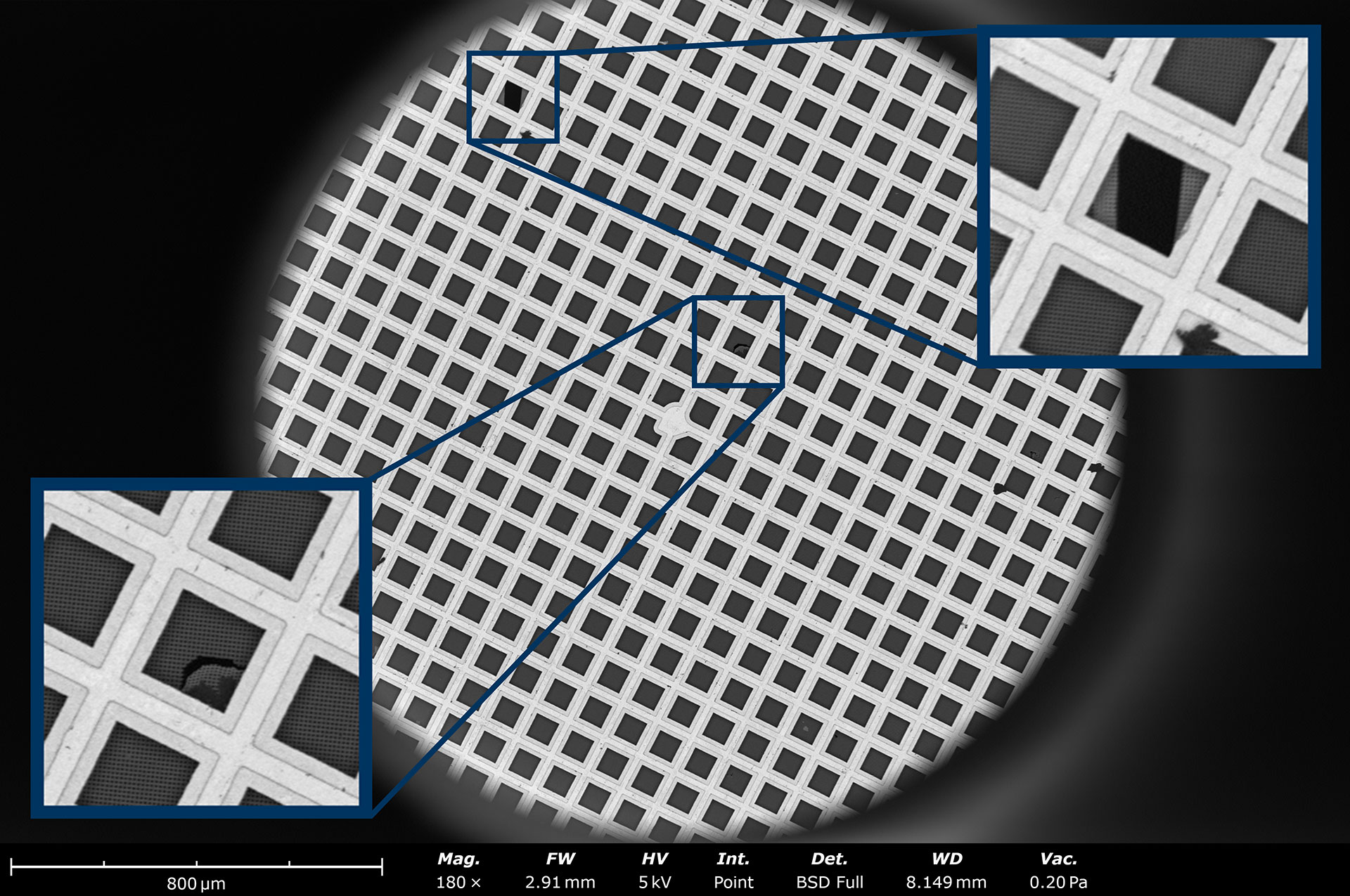
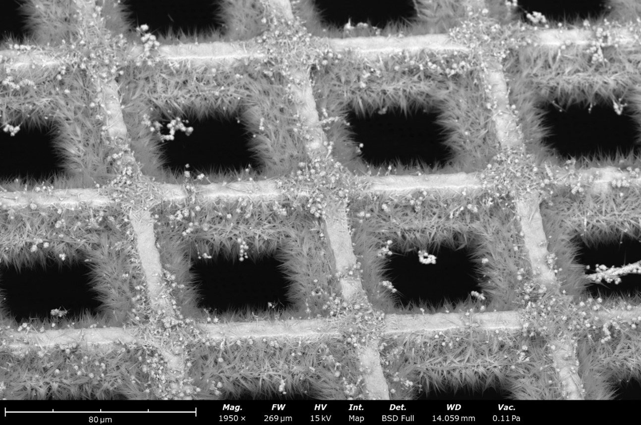
The Phenom is a high-resolution field emission SEM that is excellent at inspecting grids slated for cryo-EM. Grids used in cryo-EM have a high degree of variability. The Phenom Pharos SEM can be used to quickly screen grid quality and identify grids with broken support film, excessive bending, uneven hole size in holey films and other irregularities (Figure 5). This process takes less than a minute per grid, including the time for sample exchange. Considering that a damaged grid identified later in the cryo-EM workflow wastes hours of machine and user time, this is a highly effective yet inexpensive screening step. Additionally, for users making their own custom designed grids (Figure 6), Phenom can provide quick feedback about overall quality and coverage.
Figure 5. A quick screen of pristine cryo-EM grids with the Phenom Pharos SEM yields information regarding overall quality including square and hole coverage, broken support film, and excessive bending in minutes.
“… it can be used to help users assess homemade graphene oxide coatings. We had several SEM/EDS users come to test the Phenom. They were impressed by the ease of the intrument and the quality of the data collected. They even asked for additional access to the instrument months after they initially used it.”
– Joshua Mendez, Staff Scientist and NYSBC MEMC Manager –
Figure 6. Custom-designed grids can be assessed for quality using the Phenom backscatter or secondary electron detectors.
“… used the phenom to assess the presence and quality of graphene on a cryoEM grid. The phenom was very easy to use, and the STEM image quality and EDS tools were excellent.”
– Eugene Chua, Scientist, NYSBC –
Conclusion
The Phenom Pharos Desktop SEM/STEM provides structural biologists with multiple solutions for streamlining experimental workflows. A thorough analysis at NY Structural Biology Center demonstrated its effectiveness at screening negative stained samples for cryo-EM, showing its potential to replace expensive room-temperature TEMs. It was also shown to provide enough detail for imaging negatively stained viruses and bacteriophages, allowing their assembly state to be studied. In SEM mode, cryo-EM grids can be quickly and effectively screened for quality, contributing to a more cost-effective allocation of downstream cryo-EM resources.
Reviewed by: Cliff Mathisen1, Ingrid Koch1, Chase Buddell2, Joshua Mendez2, Daija Bobe2 and Dr. Misha Kopylov2. 1Nanoscience Instruments, 2New York Structural Biotechnology Center