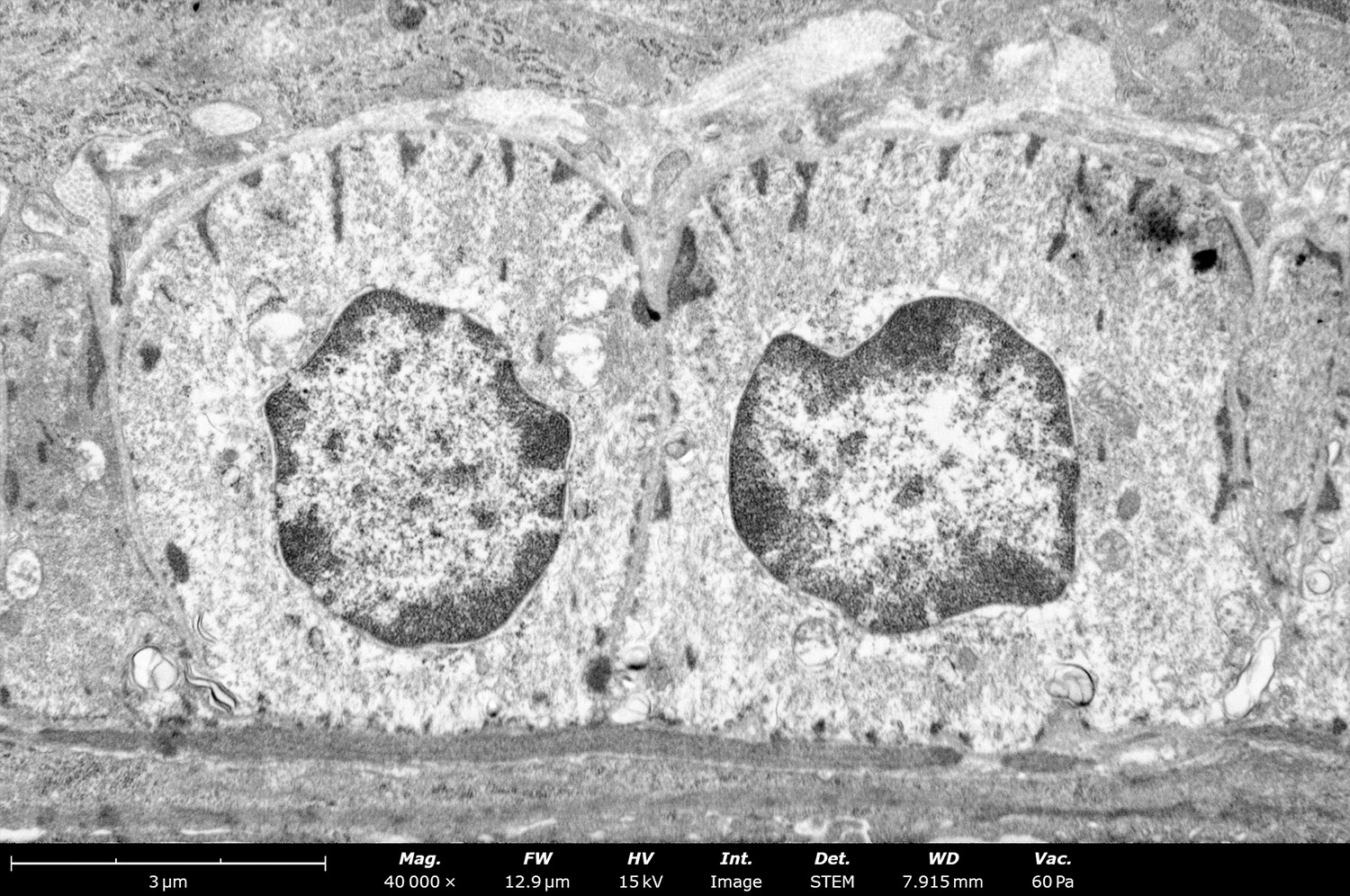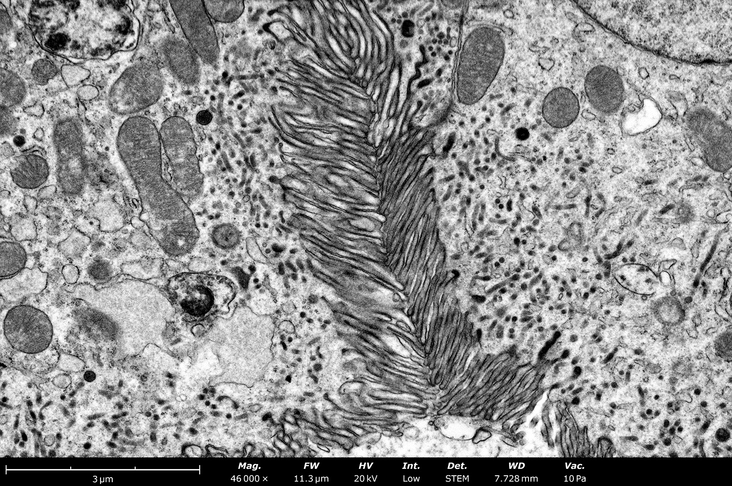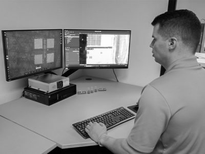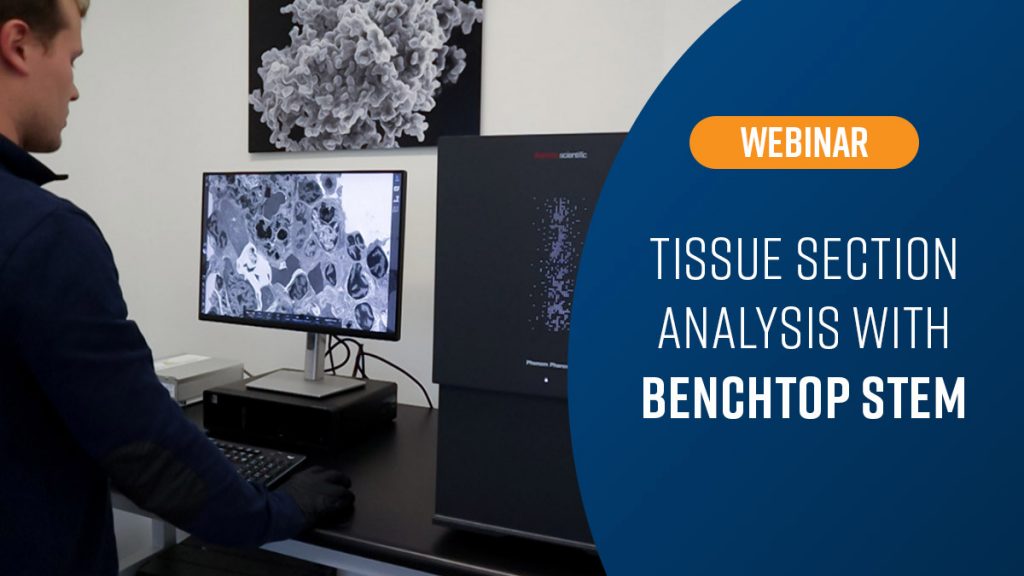

Furthering our collective understanding of biological processes is a complex, demanding endeavor. This is partly a result of the convoluted relation between macroscopic and microscopic components. One of the best tools scientists have to unravel these mysteries is one of the base human senses, sight. Viewing what goes on underneath the surface reveals a whole new world that is otherwise invisible to us. This provides an invaluable method for analyzing tissue samples, especially for disease and drug development. Electron microscopy is one of many forms of microscopic techniques used to look at tissue but it is perhaps the most powerful. Traditionally, electron microscopes were difficult to access but as technology has advanced, so has the accessibility of this technique. Desktop SEMs have become ubiquitous across many industries as a result of ease of use, small footprint, and low cost. The Phenom series of desktop SEMs, the Phenom Pharos, is a field emission gun system with a dedicated STEM holder, able to achieve the resolutions necessary to view the ultrastructures of tissue. Join us on March 20th as we dive into the benefits of electron microscopy for tissue analysis and explore how the Phenom Pharos w/STEM fits into your workflow.