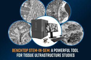
Understanding the ultrastructure of biological tissues is essential for advancing medical research, disease diagnostics, and drug development. Traditionally, histological techniques such as optical microscopy have been the standard for tissue analysis. While these methods provide valuable insights, they are often limited in resolution, making it difficult to visualize fine structural details at the nanometer scale. Transmission electron microscopy (TEM) has long been the gold standard for ultrastructural imaging,1 but conventional TEM workflows are complex, requiring dedicated infrastructure and specialized expertise.
To address these challenges, benchtop scanning transmission electron microscopy (STEM) has emerged as a powerful and accessible solution for high-resolution imaging of tissue ultrastructure. By integrating STEM capabilities within a compact scanning electron microscope (SEM), researchers can achieve nanometer-scale resolution with a simplified workflow, reducing the barriers typically associated with TEM.
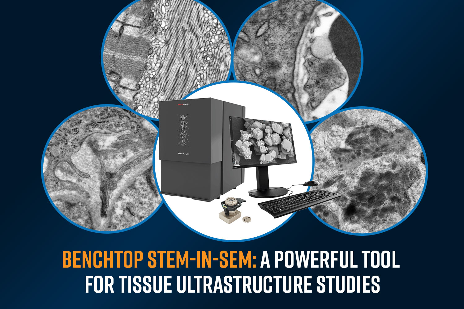
This blog explores the advantages of using STEM-in-SEM for tissue analysis, highlighting how the Phenom Pharos Desktop STEM enables effortless imaging of tissue ultrastructure. We will showcase examples of tissue section imaging acquired with the Pharos STEM, demonstrating its ability to reveal intricate cellular details with ease. Whether in pathology, neuroscience, or biomedical research, benchtop STEM opens new possibilities for high-resolution imaging without the complexity of traditional TEM workflows.
What are the benefits of STEM-in-SEM?
Traditional floor-model TEM systems are costly and resource-intensive, requiring expensive maintenance contracts, extensive user training, and long session times due to lengthy vacuum pump-down times and manual operation. As researchers demand larger datasets, there is a growing need for more accessible alternatives that still deliver high-quality imaging results.
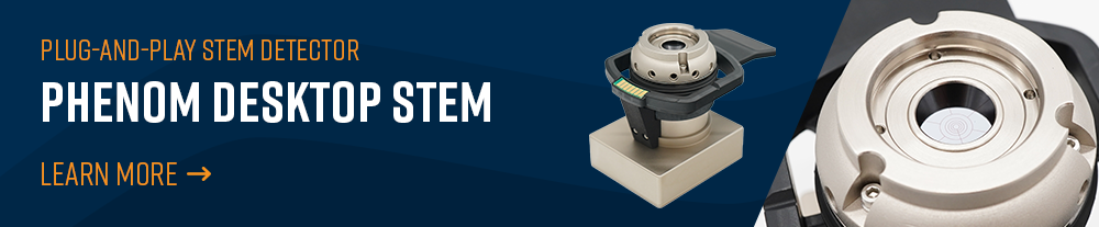
STEM-in-SEM refers to using a SEM to capture transmission electron micrographs.2 While various methods exist, the highest-quality results come from using a dedicated STEM detector (as pictured in Figure 1) rather than mirrors that redirect transmitted electrons to existing SEM detectors. In this approach, an electron-transparent sample is placed in the SEM chamber, and as the electron beam scans across it, transmitted electrons are collected by a detector positioned beneath the sample. This result is a true high-resolution image of the transmitted electrons with a high signal to noise ratio. Typically, the SEM used for STEM-in-SEM will have a field-emission electron gun, which allows it to form a focused probe at the sample surface and thus provide the best possible resolution.
Several aspects of STEM-in-SEM can improve the ultrastructural imaging workflow. These include:
- High-resolution imaging: Provides sufficient resolution for most ultrastructural applications, enabling clear visualization of subcellular details.
- Lower accelerating voltage: Enhances contrast and minimizes beam damage, making it ideal for delicate biological samples or unstained tissue sections.
- Less sensitive to sample thickness: Low voltage also improves the contrast between different materials (e.g., different tissue types), making it easier to distinguish structural details in thicker sections. This enhanced contrast allows researchers to achieve good imaging without needing ultra-thin sections
- Multimodal correlation: Users can leverage backscattered electron (BSE) and secondary electron (SE) imaging modes, as well as energy-dispersive X-ray spectroscopy (EDS) analysis, that are available when operating the microscope in SEM mode.
- Larger field of view: The STEM-in-SEM offers a larger field of view. Being able to view larger areas of the sample enables the user to find areas of interest much faster than a traditional TEM.
Using the Phenom Pharos Desktop STEM
The Phenom Pharos Desktop STEM offers unparallel accessibility to STEM-in-SEM and is exceptionally useful for imaging tissue sections at high resolutions. The compact form factor of the microscope and its vibration-resistant design allow it to be placed anywhere in the laboratory thus furthering its ease-of-use. Here are a few of the practical aspects of working with the Phenom Pharos Desktop STEM:
Fast Sample Loading
Loading samples is quick and simple (Figure 2). The sample chuck holds standard 3-mm TEM grids securely. To load, lift the copper clip, place the grid in the designated area with tweezers, and close the clip. Insert the chuck into the sample holder above the detector, then slide the holder into the chamber until the green indicator light turns on, confirming a secure load.
Using the STEM Detector
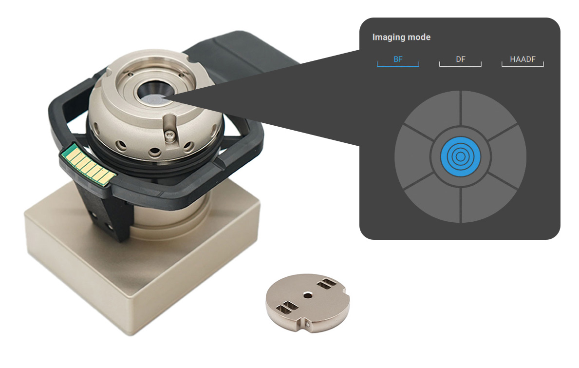
Acquiring a STEM image can be completed within a few minutes, often less than a minute of loading the sample, as shown in the video in Figure 3. Once the sample holder is inserted, the optical camera automatically displays the grid, allowing the user to select an area for imaging. In the video demonstration, the user first selects the backscattered electron detector (BSD), then switches to the STEM detector, adjusting focus and brightness for optimal quality. Switching between imaging modes is effortless as users can click preset options in the interface or customize active detector segments using the “show more” control button.
Low Vacuum Mode
Charging is one of the most consistent obstacles when imaging non-conductive samples in an electron microscope, such as thin tissue sections. As a result, the image brightness will appear saturated due to excess charge buildup on the sample surface, preventing its ultrastructure from being observed. The Phenom Pharos solves this with a low vacuum mode, which reduces charge buildup by introducing gas molecules to neutralize surface charging. As shown in Figure 4, this mode is easily activated from the main controls in the Phenom UI.
Gamma Adjustments
In STEM mode on the Phenom Pharos, gamma adjustments enhance image contrast more effectively than basic brightness and contrast controls. By fine-tuning gamma, users can accentuate differences between light and dark pixels, improving visibility of tissue ultrastructure. This is especially useful for thin tissue sections that exhibit high transmission signals, as shown in Figure 5.
Imaging Tissue Ultrastructure – Some Examples
Below are a few examples of different tissue ultrastructure observed using the Phenom Pharos Desktop STEM.
Kidney Tissue
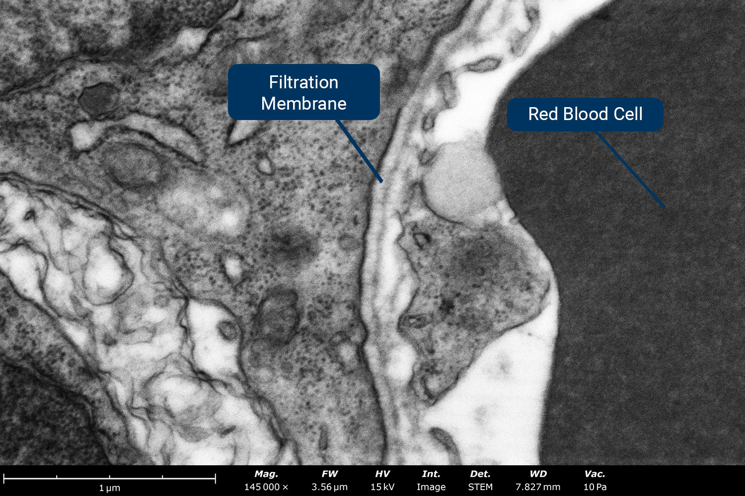
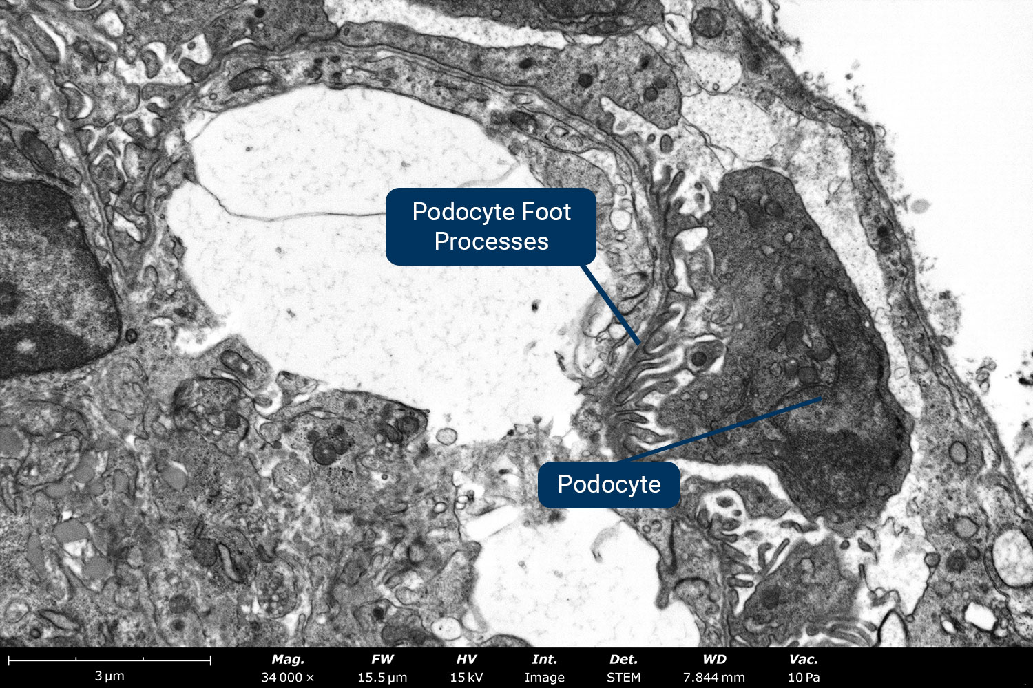
Transmission imaging provides the resolution needed to reveal key structures in kidney tissue with remarkable clarity. In Figure 6a, the filtration membrane is distinctly visible, allowing for precise thickness measurement at approximately 65 nm. Figure 6b highlights the intricate details of podocyte foot processes, showing widths of around 80 nm and varying lengths.
Liver Tissue
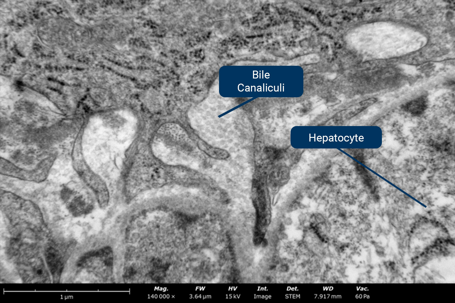
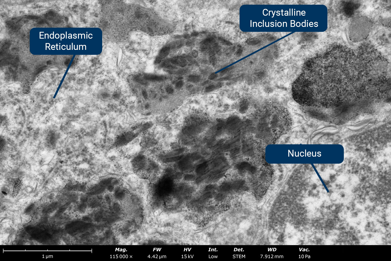
STEM imaging reveals fine structural details within liver tissue, including bile canaliculi (Figure 7a), which are the channels in-between hepatocytes that collect the bile produced from these cells, which leads to the bile ducts. At even higher magnification (Figure 7b), crystalline inclusion bodies and the endoplasmic reticulum within individual hepatocytes become distinctly visible.
Skeletal Muscle
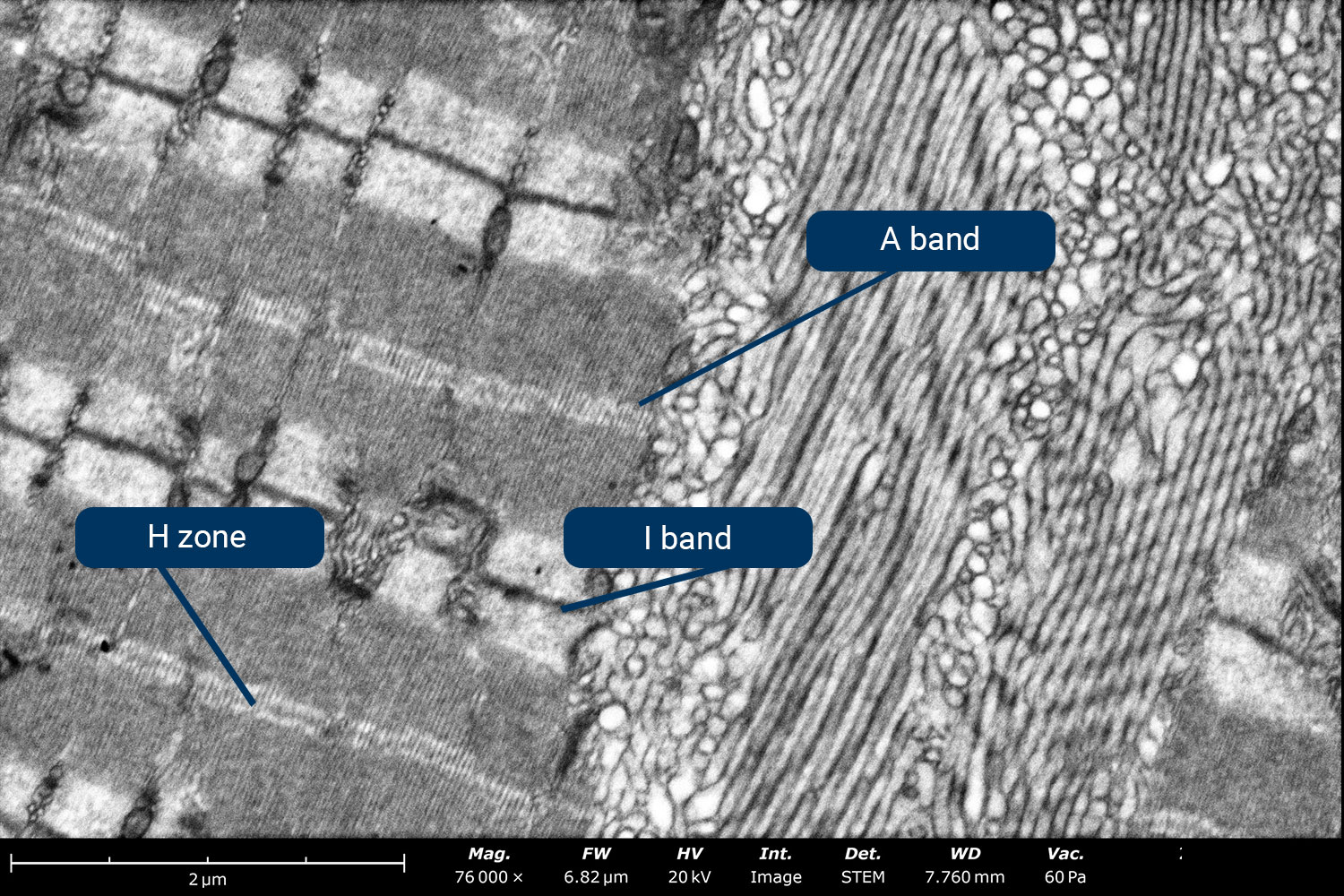
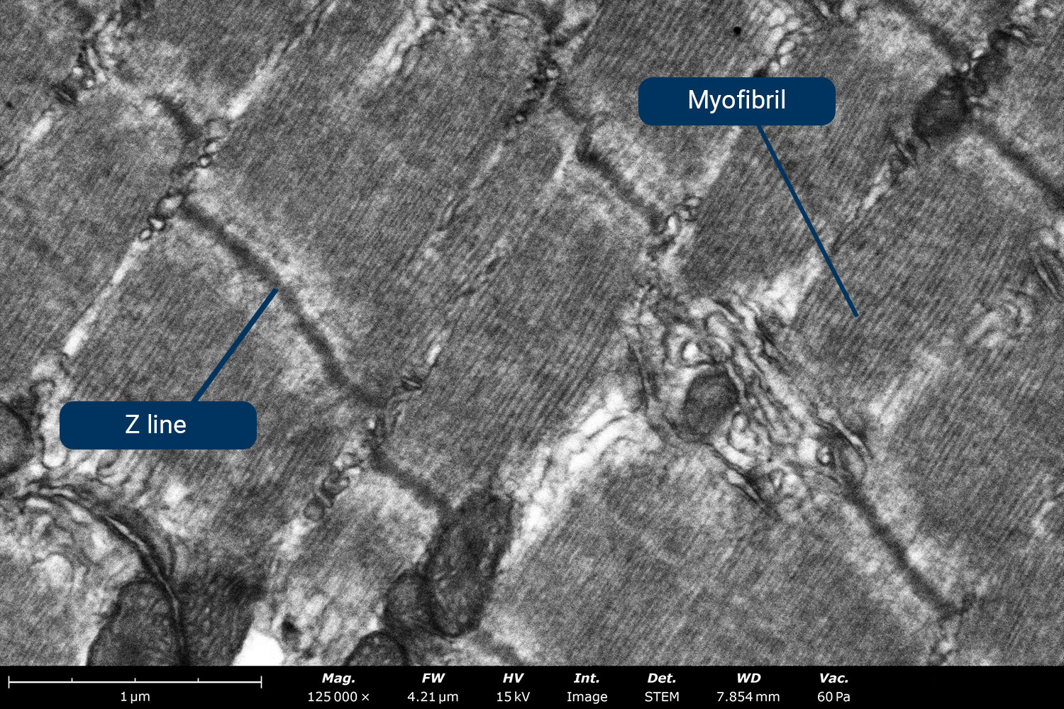
Skeletal muscle analysis using electron microscopy is crucial for understanding tissue function as well as diagnosing diseases. Figure 8a shows a BF-STEM image showing several structures within sarcomere (a structural unit of myofibril in striated muscle), including the H zone, A band, and I band. Upon observing the sarcomere at higher magnification (Figure 8b), the Z line and individual myofibrils are visible.
Optic Nerve Tissue
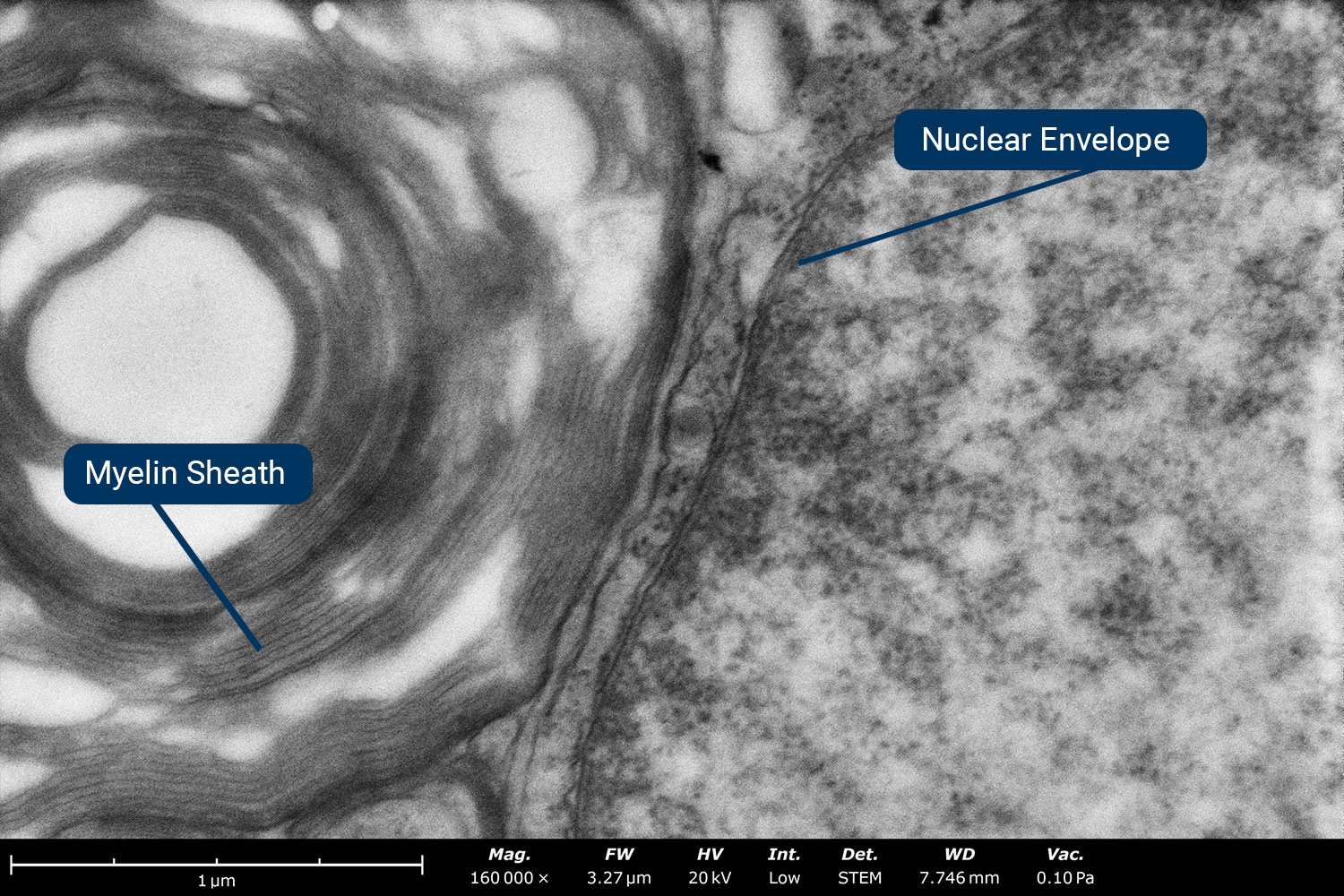
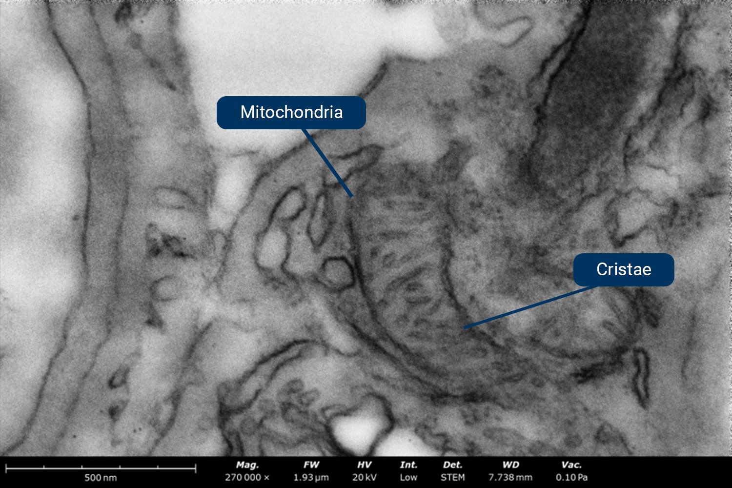
The ultrastructure of optic nerve tissue consists primarily of axons, which communicate signals from the optic nerve to the brain. Figure 9a shows a detailed image of an individual axon surrounded by myelin sheath. The myelin sheath insulates the neurons and helps them communicate effectively. Figure 9b, captured at even higher magnification, shows cristae folds within the mitochondria.
Summary
Benchtop STEM-in-SEM offers a powerful and accessible solution for imaging tissue ultrastructure while effectively overcoming the pitfalls of traditional TEM workflows. By integrating STEM capabilities within an SEM, high-resolution transmission imaging can be accomplished with a simplified workflow, reducing costs and operational complexity. The Phenom Pharos Desktop STEM streamlines tissue analysis with fast sample loading, multimodal imaging, low vacuum mode for charge reduction, and gamma adjustments for enhanced contrast. Demonstrations on kidney, liver, skeletal muscle, and optic nerve tissues showcase its ability to reveal intricate cellular structures with ease.
To explore more about this application, watch our past webinar: Tissue Section Analysis with Benchtop STEM.
References
- F. S. Sjostrand, “The Ultrastructure of Cells as Revealed by the Electron Microscope,” International Review of Cytology, vol. 5, pp. 455-533, 1956 ↩︎
- J. D. Holm and B. W. Caplins, STEM-in-SEM: Introduction to Scanning Transmission Electron Microscopy for Microelectronics Failure Analysis, ASM International, 2020. ↩︎