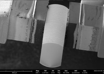
Welcome to the first scanning electron microscopy (SEM) edition of the Nanoscience Instruments newsletter, nanoNews.
In this newsletter, we will explore a range of topics relating to SEM, imaging, sample preparation methods, and data analysis tools. We hope to provide a comprehensive overview of activity in the realm of Phenom Desktop SEM, and inspire you to further explore this fascinating area of research.
Thank you for joining us on this journey of discovery.
Nanoscience Instruments Celebrates
20 Years Serving the Nano World!
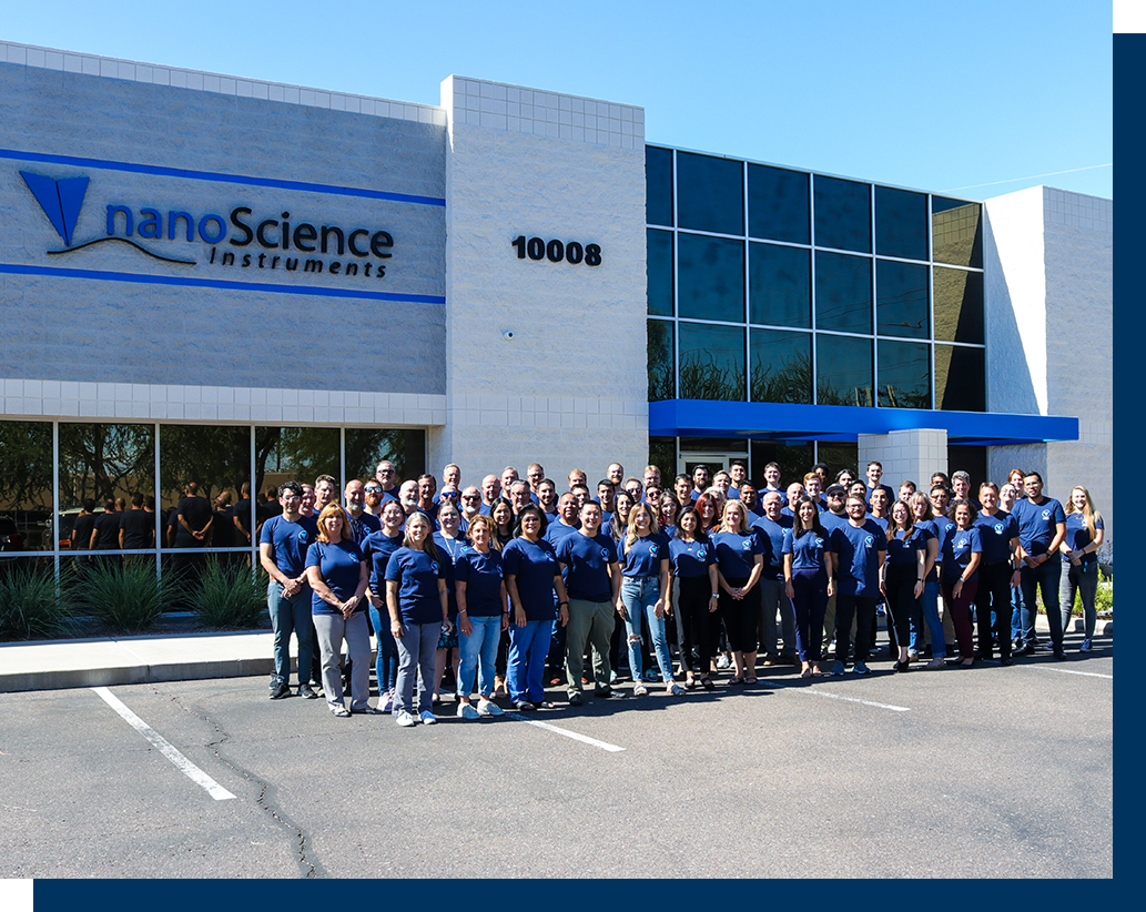
We celebrated our 20th anniversary in 2022. Find out more about our company’s history and see what our co-CEOs shared about our continued commitment to driving innovation in our recent press release:
NEWS & UPDATES

Our new website has launched! It is designed to provide you easier access to the solutions we offer for electron microscopists, as well as resource libraries that are continuously expanding with new webinars, videos, app & tech notes, and more!
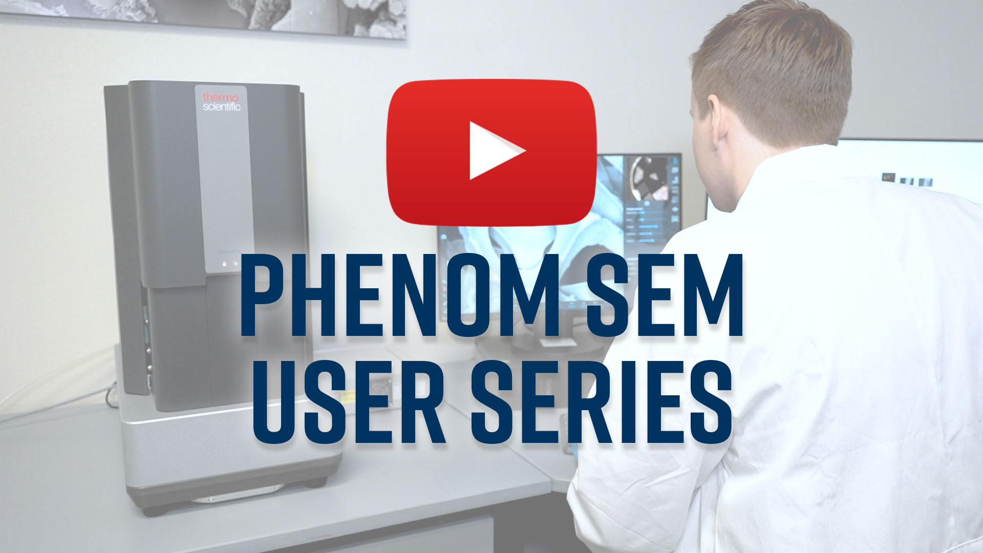
Phenom Desktop SEM User Series
New Video Playlist
We are excited to announce the launch of our new YouTube playlist, which focuses on the use of Phenom desktop scanning electron microscopes. In this series, our application scientists provide you with an in-depth look at the capabilities of Phenom, as well as tips and tricks for optimizing your imaging results. Subscribe to stay up to date!
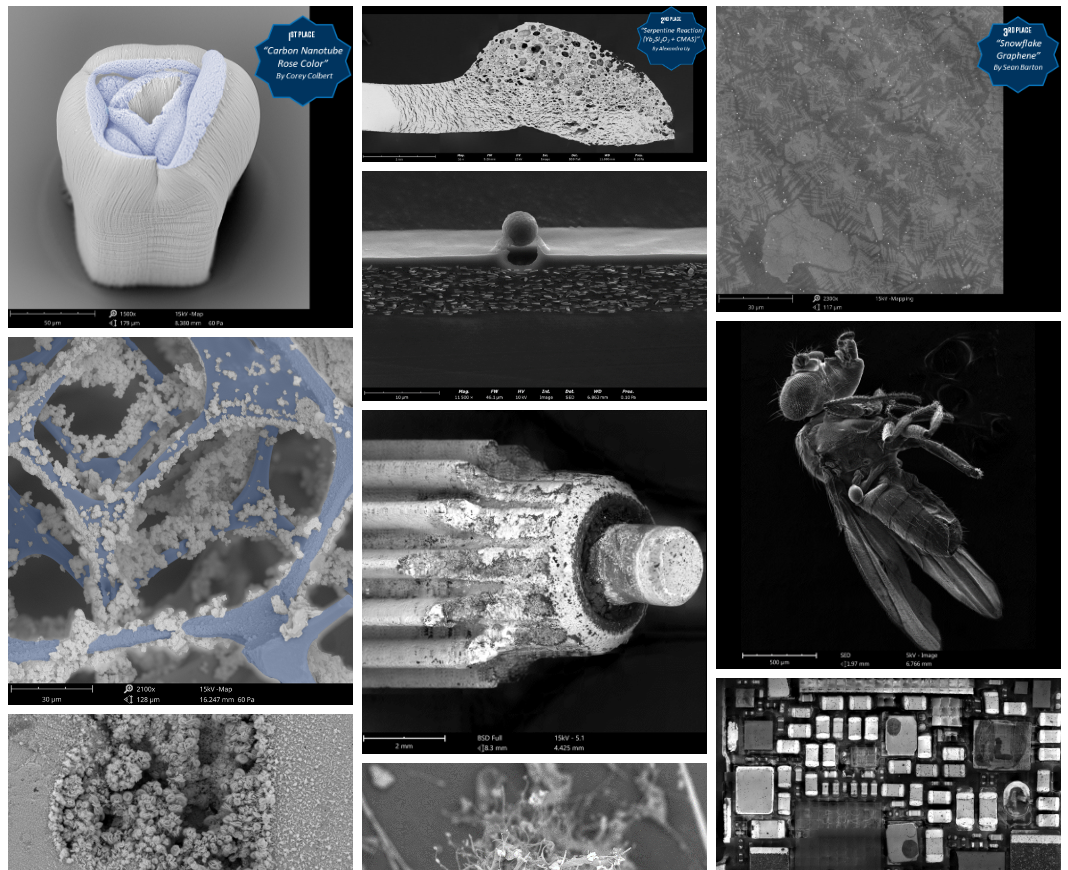
2022 Phenom Desktop SEM Image Competition
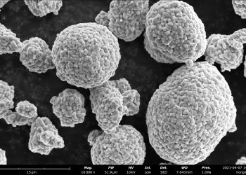
This competition aimed to recognize the extraordinary beauty and complexity of the microscopic world and to encourage our customers — scientists, researchers, and artists — to explore and share the wonders of science at a new level. The criteria used for judging included creativity, technical proficiency, informational content, and visual impact.
VIEW WEBINARS ON DEMAND
What is ParticleX? Automated Particle Analysis for Scanning Electron Microscopy
Routine and manual SEM characterization can be a tedious labor that leaves little room to perform additional analysis and reporting without consuming more time. To remedy these issues and add …
Advancement of Tissue Engineering Using Desktop SEM
Tissue engineering is a progressively advancing field focusing on biological substitutes to improve or restore functions to damaged tissues or organs in the body. Many of these substitutes are found …
Leveraging the Power of Desktop SEMs for Drug Development
Drug development requires rapid data driven decisions to address the challenges in formulation and keep pace with the demand for higher therapeutic effectiveness. As drug development increasingly focuses on the …
Morphological and Compositional Analysis of Battery Materials
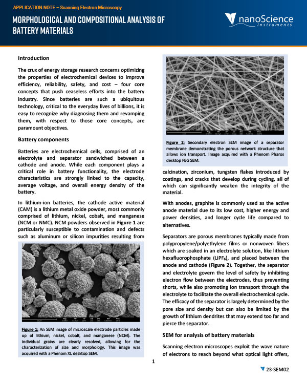
With the increasing demand for clean and sustainable energy, there is an ongoing need for more efficient and reliable energy storage systems. At the foundation of these systems, R&D and …
If SEM, Then Script: Python Coding for Scanning Electron Microscopes
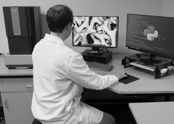
This upcoming webinar will showcase the innovative application of Python programming to control the Phenom Desktop SEM. Attendees will learn how Python can be used to automate the SEM imaging …
APPLICATION NOTE & BLOG UPDATES
Below you’ll find the newest written documents that showcase the limitless capabilities of SEM technology. We hope you’ll gain a deeper appreciation for the power of SEM and its role in advancing scientific understanding across a wide range of fields, from biology and medicine to materials science and beyond.
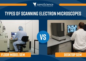
Desktop SEM vs Floor Model SEM: A Comparison
In the realms of scientific industry and research, scanning electron microscopes are among the most common imaging tools for studying the microstructures of materials. Microscopists employ SEMs to greatly expand …
Why Use An SEM in Battery Research?
The functional properties of a battery, not limited to just its performance, are inherently dependent upon the microstructure and surface morphology of electrodes and separators. Scanning electron microscopy is a …
Which Electron Source is Best?
The electron source is one of the most important components of a scanning electron microscope (SEM) and is a major factor in determining its maximum analytical performance. There are three …
How to Combat Electric Charge Buildup in Scanning Electron Microscopy
The fundamental unit of electric charge is the electron, an elementary particle that can be used in the domain of microscopy to see beyond the diffraction limit of optical light …
SERVICE & SUPPORT
Are you aware that we provide both on-site service and remote access software assistance for your SEM maintenance and repair needs?
For inquiries regarding service or maintenance, please contact our dedicated service engineer team at service@nanoscience.com.
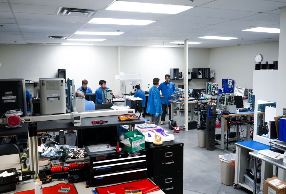
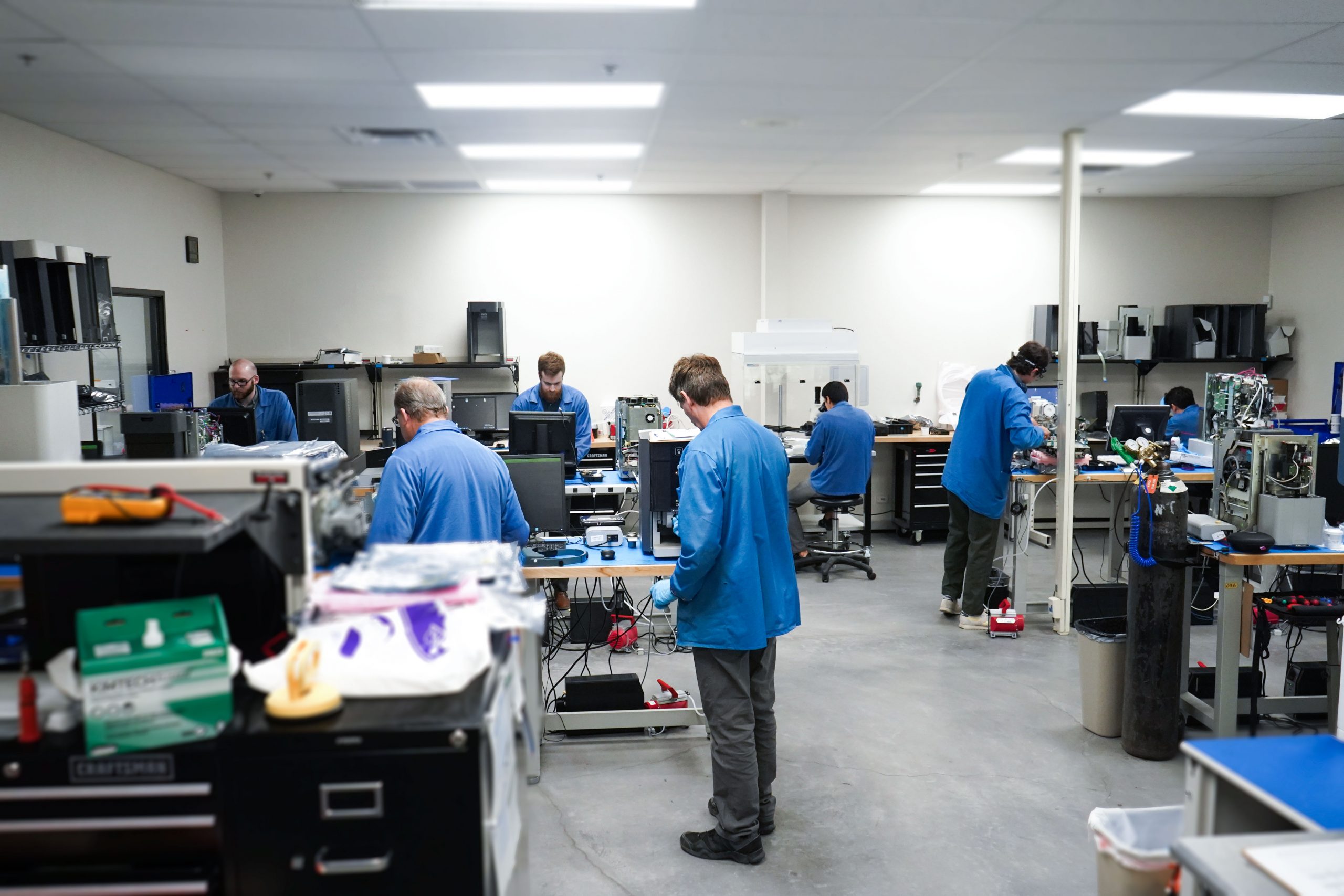
At Nanoscience Instruments, we take great care in ensuring that your Phenom Desktop SEM systems are operating at their optimal performance levels, while also providing exceptional customer service. To this end, we have a curated team of highly skilled and experienced service engineers who are well-versed in all aspects of SEM maintenance and repair.
Whether you require preventative maintenance, routine checkups, or a diagnosis, our team is on hand to provide on-site service or connect with you through remote access software for troubleshooting and repair support. We understand that downtime can be costly, so our goal is to minimize it by providing efficient and effective solutions.
But our service team is more than just technical expertise. They pride themselves on cultivating positive working relationships with system users. We believe that this not only facilitates effective communication, but also builds a strong foundation of trust between our team and our customers.
CUSTOMER SHOWCASE
In this section of our newsletter, we shine a spotlight on our valued customers who are utilizing scanning electron microscopes in their respective fields. From researchers exploring the depths of nanotechnology to engineers developing cutting-edge materials and devices, these individuals and research teams are pushing the boundaries of science with the help of SEMs.
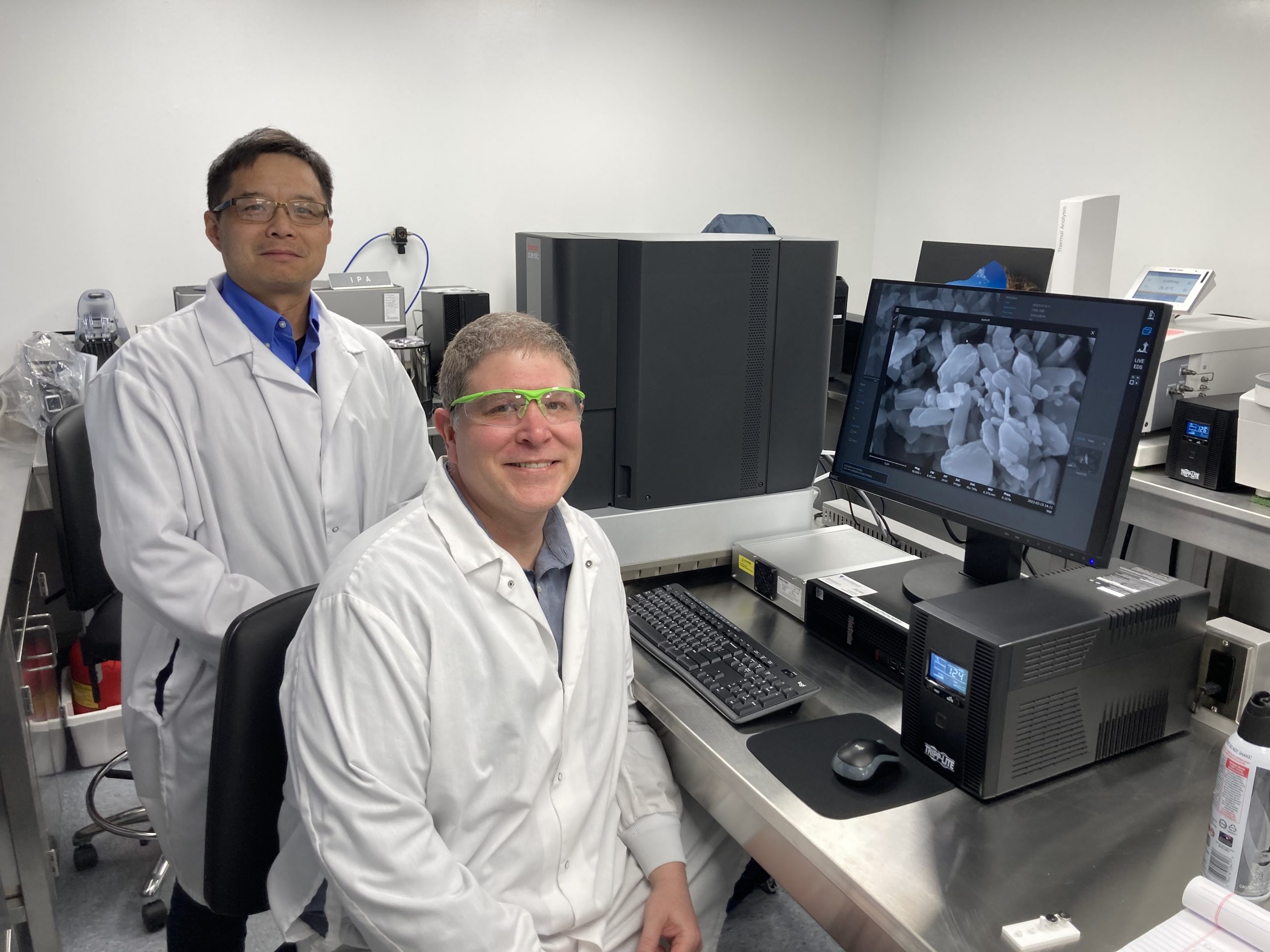
Microsize
Phenom XL
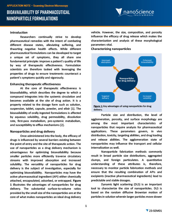
Microsize plans to delve deeper into the properties of their APIs (active pharmaceutical ingredients) and excipients by closely examining particle properties. Monitoring various aspects of their powders like agglomeration, wetting, bioavailability, and so on, allows Microsize to maintain the highest standards of quality control.
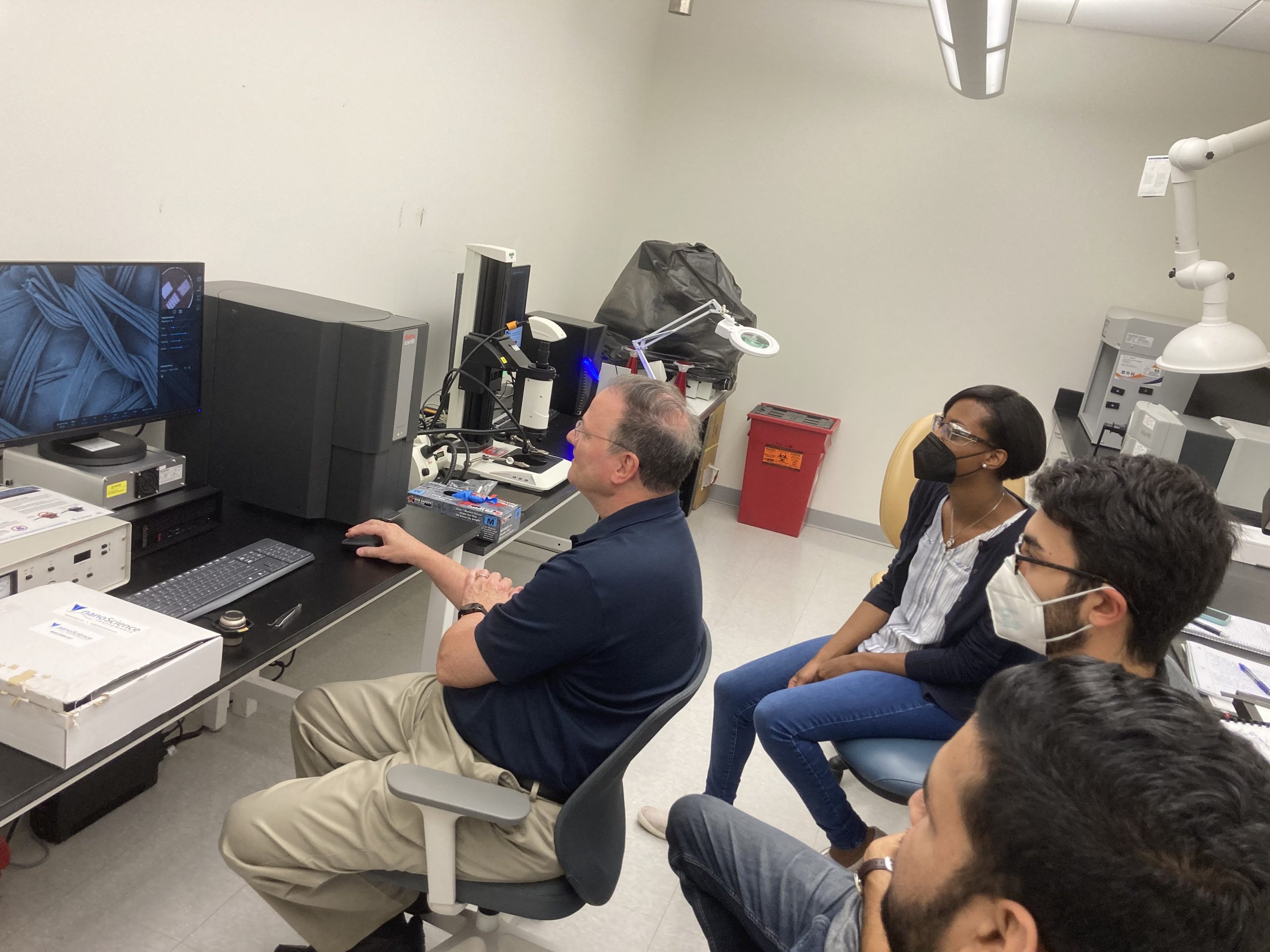
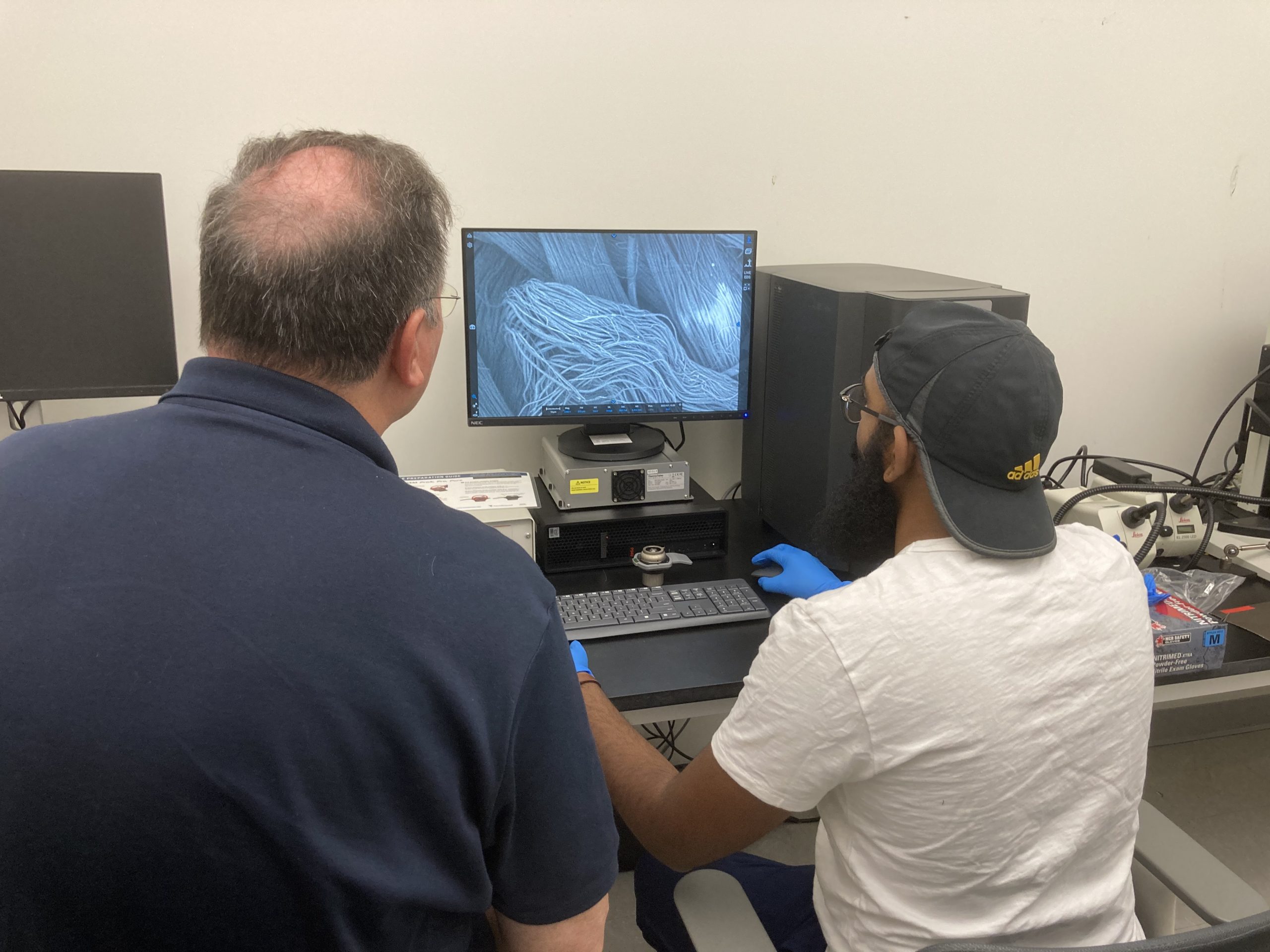
Soliyarn
Phenom XL
Soliyarn’s scientists are dedicated to developing and manufacturing high-performance textiles with conductive and semiconductive properties. By using unique coating and weaving methods, they develop heated garments, sensing textiles, and repellent fabrics. Advanced microscopy and spectroscopy techniques are therefore necessary to analyze and characterize their materials, allowing them to maintain high standards of quality and performance in their products.
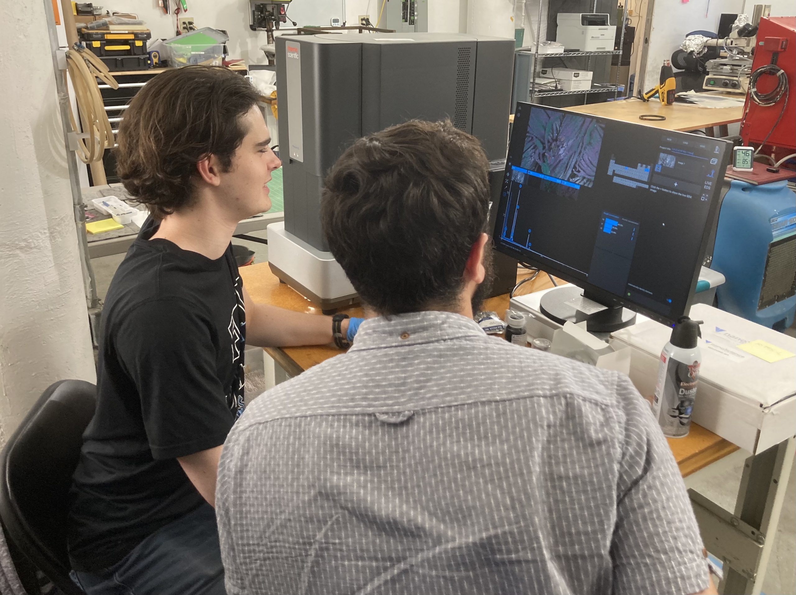
Modern Meadow
Phenom ProX
Modern Meadow specializes in creating biofabricated materials and uses SEM to analyze the surface structure and composition of their materials at high magnification. By examining the microscopic properties of their biofabricated materials, they can better understand their performance and identify areas for improvement.
In addition to advanced microscopy techniques, elemental analysis by way of energy dispersive X-ray spectroscopy (EDS) is crucial for ensuring the quality, consistency, and sustainability of their products.
SEM PUBLICATIONS
The publication section showcases the latest and greatest articles about SEM. We provide you with a curated selection of articles that demonstrate the diverse applications of SEM and how these techniques are critical tools that allow us to explore and understand the intricate details of our world.
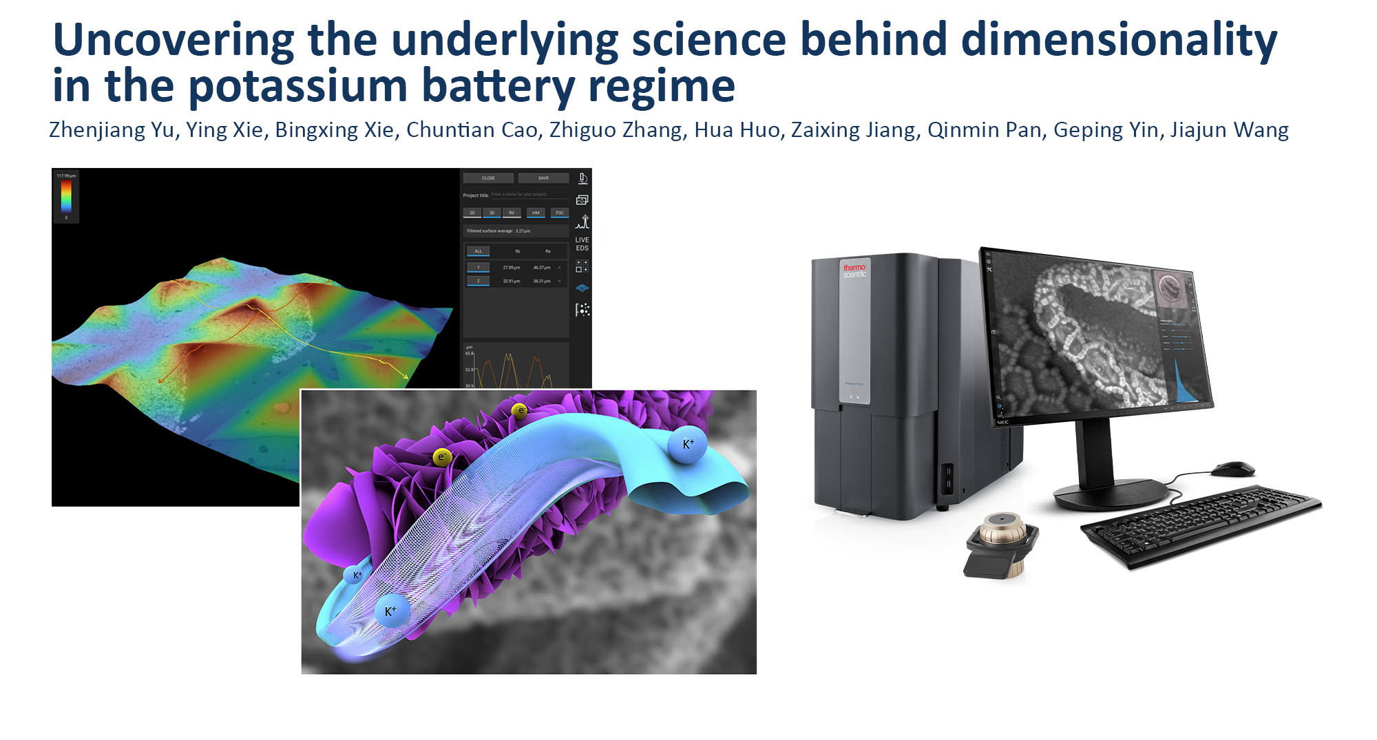
Uncovering the underlying science behind dimensionality in the potassium battery regime
Discover Further Reads…
- Analysis of ultrastructure and microstructure of blackbird (Turdus merula) and song thrush (Turdus philomelos) eggshell by scanning electron microscopy and X-ray computed microtomography: https://www.nature.com/articles/s41598-022-16033-5
- Influence of cold atmospheric plasma on dental implant materials – an in vitro analysis: https://link.springer.com/article/10.1007/s00784-021-04277-w


