PHENOM DESKTOP SEM
Electron Detectors
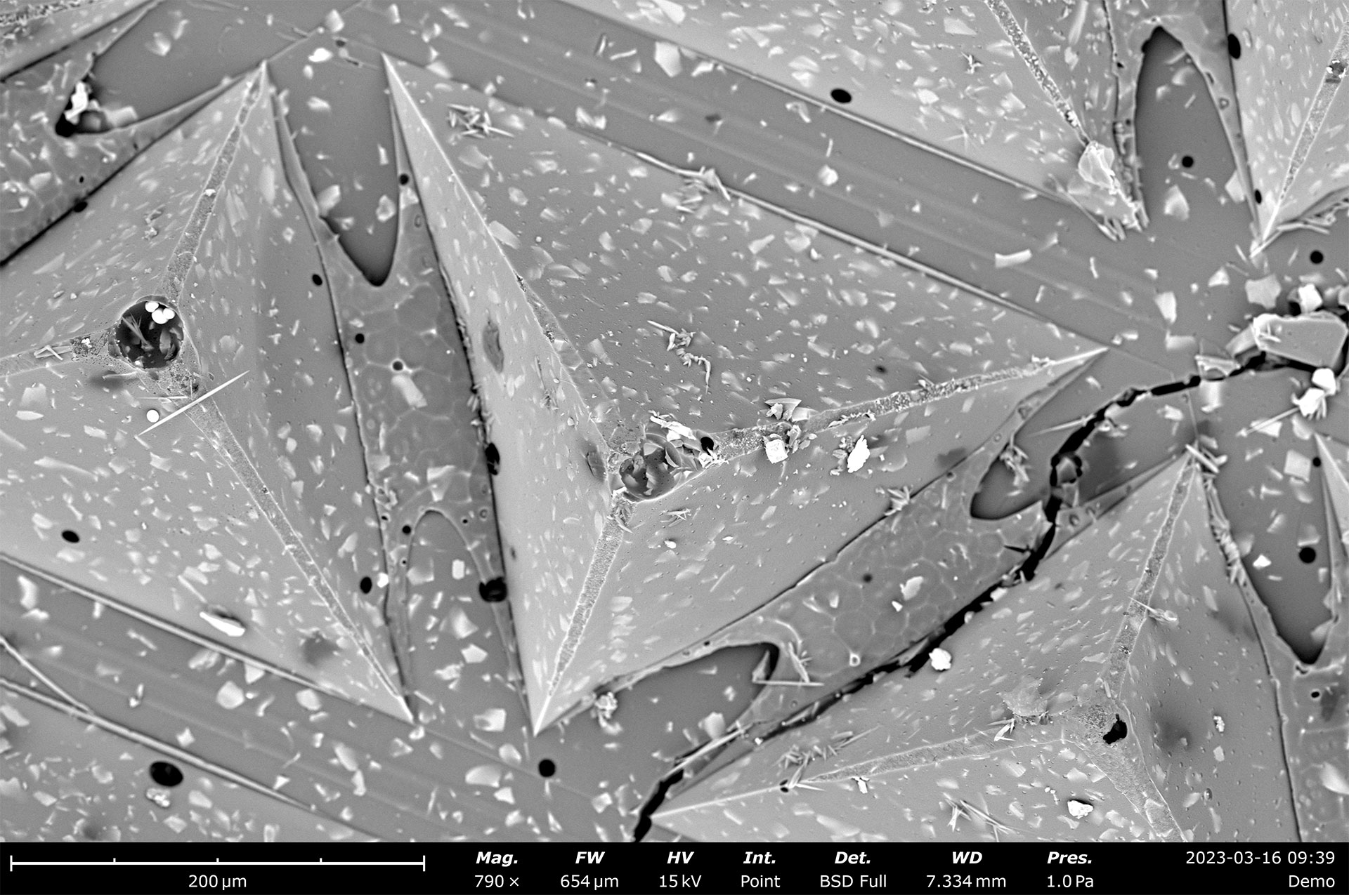
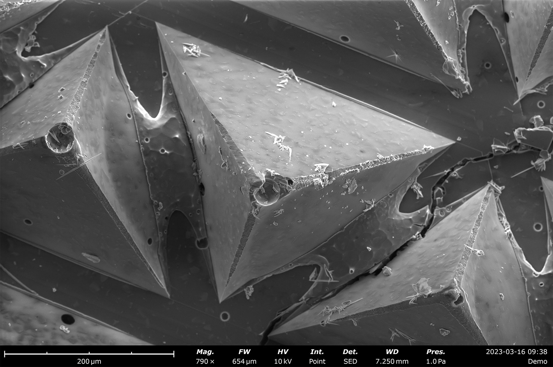
The Phenom Backscattered Electron Detector (BSD) and Phenom Secondary Electron Detector (SED) are responsible for detecting electrons resulting from interactions between the electron beam and sample surface. The two detectors collect different signals that convey varying information about atomic numbers and surface topography.
Fast Imaging
High sensitivity paired with the fastest vent-load cycle provides an average time-to-image of under 40 seconds
Mixed Mode
Built-in signal mixing allows you to simultaneously view BSD and SED signals in a singular composite image
Surface Sensitivity
Topographic imaging using an optional SED, or take advantage of the segmented BSD in topographic mode
Talk to an Instrumentation Specialist Today!
Overview of Phenom Electron Detectors
The Phenom BSD provides high resolution images that convey elemental contrast information. This highly sensitive detector is standard to every Phenom Desktop SEM and is configured to provide fast, high-quality imaging that allows you to readily identify the microstructure of a sample. The detector operates well at any magnification and vacuum level and an optional topographic mode makes use of the four-segment configuration of the sensors to provide qualitative and three-dimensional visualizations of surface topography.
The Phenom SED collects electrons emitted from the sample surface and thus provides crisp and high-resolution surface-sensitive imaging. Images generated with the SED convey surface topographical information and generally have higher resolution as a result of the smaller beam-sample interaction volume. Therefore, SED imaging is a great complement to BSD imaging overall.
Phenom Electron Detectors
Product Features
Compositional imaging mode
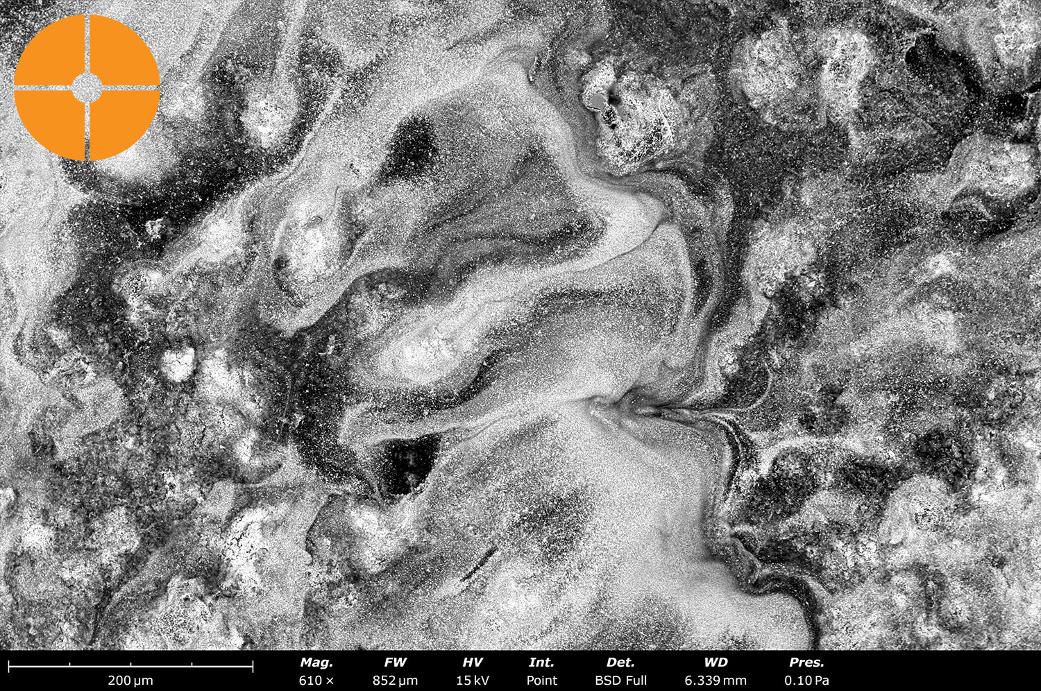
The standard backscattered electron detector (BSD) is a four-segment detector that provides high signal-to-noise images with compositional contrast. BSD imaging provides rapid, high-resolution images that reveal a sample’s microstructure in exceptional detail, enabling clear differentiation between regions of varying elemental composition. This versatile imaging mode is valuable across a wide range of applications, from materials science to failure analysis, offering fast and reliable insights into complex sample structures.
Topographic imaging mode
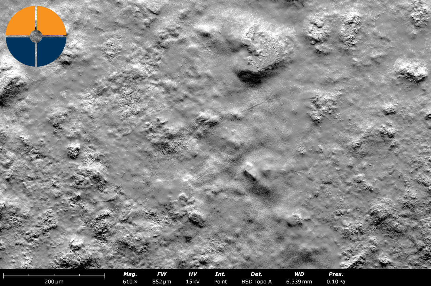
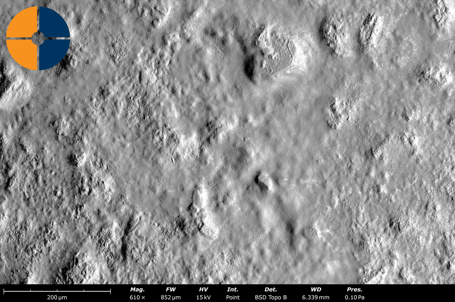
The topographic imaging modes, Topo A and Topo B, enhance surface analysis by displaying the BSE signal difference between the upper/lower and left/right halves of the backscatter detector (BSD). These modes provide a unique way to visualize surface topography, offering qualitative insights into height variations and surface features that may not be apparent in standard BSE images.
3D roughness reconstruction
The 3DRR technique generates a 3D surface map, enabling detailed roughness measurements and advanced surface analysis. By applying a “shape from shading” algorithm, it calculates relative surface heights based on signal variations from the four quadrants of the backscatter detector (BSD). The results are displayed as a heat map, providing more intuitive and detailed topographical information compared to standard grayscale images.

3D Roughness Reconstruction of fuel cell surface, showing areal average roughness (Sa) and line-profile roughness (Ra).
Secondary electron imaging
Secondary electrons provide detailed surface information because they emerge from an interaction volume that is confined near the sample surface. SED images provide excellent topographic contrast at finer resolution than what can be achieved using a BSD.
Phenom Electron Detectors
Product Knowledgebase
Webinar
Practical SEM and Ion Mill Applications for Semiconductor R&D to Production
Scanning Electron Microscopes (SEMs) and Cross-sectioning/Polishing tools play a vital rol…
Blog
Optical Microscopy vs SEM for Technical Cleanliness Analysis
The performance of sensitive systems such as engines, hydraulics, electronics, and medical…
White Paper
Understanding Nickel Manganese Cobalt (NMC) Precursor Cathode Active Material (pCAM)
As the battery industry strives for more efficient and environmentally friendly solutions…


